Home » Without Label » Back Muscles Anatomy / Back Strains And Sprains - The muscles of the lower back, including the erector spinae and quadratus lumborum muscles, contract to extend and laterally bend the vertebral column.
Back Muscles Anatomy / Back Strains And Sprains - The muscles of the lower back, including the erector spinae and quadratus lumborum muscles, contract to extend and laterally bend the vertebral column.
Back Muscles Anatomy / Back Strains And Sprains - The muscles of the lower back, including the erector spinae and quadratus lumborum muscles, contract to extend and laterally bend the vertebral column.. Superficial back muscles, intermediate back muscles and intrinsic back muscles.the intrinsic muscles are named as such because their embryological development begins in the back, oppose to the superficial and intermediate back muscles which develop elsewhere and are therefore classed as extrinsic muscles. Includes latissimus dorsi, the trapezius, levator scapulae and the rhomboids. Muscle origin insertion action innervation artery notes; Back pain is common and might be caused by a problem with a muscle. These muscles provide posture and stability to the body by holding the vertebral column erect and adjusting the position of the body to maintain balance.
Balance the weight of your head on top of your spine evenly distribute weights from your upper body into the lower extremities Muscles labeled front and back These muscles provide posture and stability to the body by holding the vertebral column erect and adjusting the position of the body to maintain balance. Able to move the upper limb as they originate at the vertebral column and insert onto either the clavicle, scapula or humerus. The deep muscles develop in the back called intrinsic muscles.
How To Draw The Torso Back View from static.wixstatic.com Human musculature bodybuilding infographic muscular system vector human anatomy back muscle anatomy bicep male muscular anatomy human body anatomy female female anatomy muscle hamstrings muscle. They start at the top of the neck and go down to the tailbone. Extends and laterally bends the neck and head, rotates head to the same side: Muscle anatomy triceps 12 photos of the muscle anatomy triceps anatomy of triceps muscle, biceps triceps muscle anatomy, muscle anatomy triceps, triceps muscle anatomy mri, human muscles, anatomy of triceps muscle, biceps triceps muscle anatomy, muscle anatomy triceps, triceps muscle anatomy mri. The back muscles are divided into two large groups: The deep muscles develop in the back called intrinsic muscles. The lower back (where most back pain occurs) includes the five vertebrae in the lumbar region and supports much of the weight of the upper body. Back pain is one of the most common kinds of pain for adults, and muscle strains are the most common type of back pain.
The muscles of the back can be arranged into 3 categories based on their location:
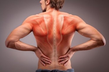
All about the back muscles the back anatomy includes the latissimus dorsi, trapezius, erector spinae, rhomboid, and the teres major. Superficial muscles of the back are located directly deep towards the skin along with superficial fascia.they are occasionally called the appendicular group as these muscles are mainly associated with activities of the appendicular skeleton. The lower back (where most back pain occurs) includes the five vertebrae in the lumbar region and supports much of the weight of the upper body. These structures work together to support the body, enable a range of movements, and send messages from the brain to the. These muscles give height and breadth to back development. Back pain is common and might be caused by a problem with a muscle. This curve, called lordosis, helps to: The back muscles are divided into two large groups: Muscle anatomy triceps 12 photos of the muscle anatomy triceps anatomy of triceps muscle, biceps triceps muscle anatomy, muscle anatomy triceps, triceps muscle anatomy mri, human muscles, anatomy of triceps muscle, biceps triceps muscle anatomy, muscle anatomy triceps, triceps muscle anatomy mri. Extends and laterally bends the neck and head, rotates head to the same side: The superior part of the appendicular skeleton that includes clavicle, scapula, and humerus, is attached to the axial skeleton that consists of skull. The human spine is composed of 4 sections of vertebrae. The muscles of the lower back, including the erector spinae and quadratus lumborum muscles, contract to extend and laterally bend the vertebral column.
Able to move the upper limb as they originate at the vertebral column and insert onto either the clavicle, scapula or humerus. The intrinsic back muscles are found deeper to the extrinsic muscles, separated from them by the thoracolumbar fascia. This curve, called lordosis, helps to: Anatomy of the back muscles the latissimus dorsi muscles (also known as the lats) are the largest muscles of the back. The superficial back muscles are situated underneath the skin and superficial fascia.
The Anatomy Of The Back Muscles Step To Health from steptohealth.com Superficial back muscles, intermediate back muscles and intrinsic back muscles.the intrinsic muscles are named as such because their embryological development begins in the back, oppose to the superficial and intermediate back muscles which develop elsewhere and are therefore classed as extrinsic muscles. Leaning back to straight vertical and all points in between. This curve, called lordosis, helps to: 1 your spine in this region has a natural inward curve. (2017, elsevier) should be consulted. Muscles of the lumbar spine. Includes latissimus dorsi, the trapezius, levator scapulae and the rhomboids. The muscles of the back can be arranged into 3 categories based on their location:
See back muscle anatomy stock video clips.
The back muscles are divided into two large groups: Back muscles anatomy the surface muscles of the upper back include the trapezius muscles (traps) and posterior deltoids. Browse 3,579 back muscle anatomy stock photos and images available, or search for pelvic bone or lymphatic system to find more great stock photos and pictures. All about the back muscles the back anatomy includes the latissimus dorsi, trapezius, erector spinae, rhomboid, and the teres major. Superficial back muscles, intermediate back muscles and intrinsic back muscles.the intrinsic muscles are named as such because their embryological development begins in the back, oppose to the superficial and intermediate back muscles which develop elsewhere and are therefore classed as extrinsic muscles. They provide movements of the spine , stability to the trunk, as well as the coordination between the movements of the limbs and trunk. Muscle origin insertion action innervation artery notes; Leaning back to straight vertical and all points in between. The back muscles are anatomically layered into superficial (extrinsic) and deep (intrinsic) muscles. On this page, you'll learn about each of these muscles, their locations and functional anatomy. Understanding lower back anatomy is key to understanding the root of lower back and hip pain. The lower back (where most back pain occurs) includes the five vertebrae in the lumbar region and supports much of the weight of the upper body. What are the lower back muscles and their anatomy?
Mastoid process and lateral end of the superior nuchal line: The human spine is composed of 4 sections of vertebrae. Muscle origin insertion action innervation artery notes; The muscles of the back can be arranged into 3 categories based on their location: Anatomy chart courtesy of fcit the latissimus dorsi muscles (also known as the lats) are the largest muscles of the back.
How To Train Your Lower Back Muscles Exercises Workout Strengthlog from i2.wp.com Three types of back muscles that help the spine function are extensors, flexors and obliques. Back pain is common and might be caused by a problem with a muscle. This blog post article is an overview of the muscles of the lumbar spine of the trunk. The deep muscles develop in the back called intrinsic muscles. Muscles labeled front and back Understanding lower back anatomy is key to understanding the root of lower back and hip pain. The muscles, bones, ligaments, and tendons in the back can all be injured and cause back pain. The human spine is composed of 4 sections of vertebrae.
Superficial muscles of the back are located directly deep towards the skin along with superficial fascia.they are occasionally called the appendicular group as these muscles are mainly associated with activities of the appendicular skeleton.
Anatomy of the back muscles the latissimus dorsi muscles (also known as the lats) are the largest muscles of the back. Browse 3,579 back muscle anatomy stock photos and images available, or search for pelvic bone or lymphatic system to find more great stock photos and pictures. The back muscles are divided into two large groups: See back muscle anatomy stock video clips. Leaning back to straight vertical and all points in between. Muscles labeled front and back The intrinsic back muscles are found deeper to the extrinsic muscles, separated from them by the thoracolumbar fascia. Anatomy chart courtesy of fcit the latissimus dorsi muscles (also known as the lats) are the largest muscles of the back. Includes latissimus dorsi, the trapezius, levator scapulae and the rhomboids. Muscle or ligament strains can occur from repeated use of the muscles, or from improperly or awkwardly lifting heavy objects. Back muscles anatomy the surface muscles of the upper back include the trapezius muscles (traps) and posterior deltoids. The extrinsic back muscles are located in the back, but act to produce movements of the shoulder and assist respiration. These muscles include the large paired muscles in the lower back, called erector spinae, which help hold up the spine, and gluteal muscles.